Blogger Widgets

...

TRANSLATE AS YOU LIKE

Showing posts with label Hypertension. Show all posts

May 27, 2015

Feed your Brain with ECG Today

Many people think electrocardiograph (thusly called ECG or EKG) a tough talk to understand and interpret. This is not such a huge task or need to do specialization in cardiology. This is just cardiology, simple biological pipes and wires. ECGs are simpler still, being a moving real time map of the “wires” part and how they conduct electricity.  Reading EKGs only seems hard because most cardiologists use long (expensive) words.
Most healthy hearts pump blood through the body when stimulated by an electrical signal that travels along predetermined pathways.This causes the cardiac cells to contract in just the right order, resulting in a magical four chambered pumping action. 
An EKG is a graph tracing the strength and direction of this electrical signal. Leads equipped with conductive goo are placed on different parts of the body allowing a view of the heart from different angles. If the electrical activity of the heart at any given moment is traveling toward the lead being viewed, the line on the graph goes up (positive deflection).  If the electrical activity is traveling away from the lead, the line goes down (negative deflection). This graph is being traced by a stylus on a moving piece of graph paper. In a normal healthy heart, an ECG representing one complete heartbeat looks about like this :
That first petite little hump, affectionately called the P wave, represents the electrical signal that starts in a group of cells called the Sinoatrial Node. This signal then travels through the atria (the smaller and upper two chambers of the heart) causing them to contract and push blood in to the larger and more powerful ventricles below.
The “PR Interval” segment represents a delay in the signal at another grouping of cells called the AtrioVentricular Node.This delay allows time for the atria to completely deliver their bounty into the Ventricles. With perfect timing this signal continues through the Bundle of His.The signal splits and speeds along down the left and right bundle branches, making its way to the Purkinje fibers and turning north again.This stimulates those Ventricular beefcakes to contract and deliver their payload to the lungs and body (if hearts had biceps, the left ventricle would be the proverbial “gun show”….it’s such a glory hog!).
The journey causing this second contraction through the ventricles is represented by the QRS portion of the ECG. The larger T wave which then finishes off our heartbeat is the repolarization of the ventricles.I know what you’re thinking, either “what in the what now goes where?” or hopefully, “what wave represents the repolarization of the atria”? Well, the repolarization of the atria is buried in the larger signal of the QRS and therefore not visible on the graph.
This pattern is called normal sinus rhythm.  It is the basic ECG of any normal healthy heart. Naturally, there are variations of normal within the healthy population. For example, my boyfriend is very fit and has a *huge* R wave (hands off his big left ventricle ladies, it’s all mine! And don’t get me started on his early repolarization…and no…being early in this case is definitely not a bad thing). However, anything outside of the normal range is analyzed along with the patient’s symptoms to create a working diagnosis.There are several types of common abnormalities. A PR interval that is too long is called a first degree block. A QRS that takes longer than .12 seconds is likely to be caused by a delay in one or both bundle branches, called a bundle branch block. A complete lack of P waves, and in their place a squiggly line, combined with an irregular heartbeat is likely to be atrial fibrillation. What really gets a Paramedic or ER doctor excited is when they see an elevation of the ST segment in a few consecutive leads.This is referred to as an ST elevation myocardial infarction (heart attack) and generally results in a speedy trip to the catheterization. 
Feeding points:
1) Each small box on the modern ECG strip represents 0.04 seconds on the horizontal axis.
2) ECG paper is calibrated to move at 25mm per second past the stylus.
3) The ECG was invented by the Dutch Dr. Willhelm Einthoven in 1903, for which he received the Nobel Prize in 1924. More impressively, he also had a triangle named after him.
4) It is very rare, but possible for an ECG to show a flat line, called asystole, when in fact the heart is still beating and producing a pulse.  It is because of this that it’s common practice to confirm death in a patient by looking for asystole in more than one lead.
5) Conversely it is also possible, and more common, to have an ECG show heart activity, and even normal sinus rhythm, after a person has died and their heart is no longer pumping blood. This is called a PEA, pulseless electrical activity, and shows what is left of the heart’s intact electrical system after the muscle itself has failed.
I hope you understand well. 

Thanking you
Dr.Akshaya Srikanth B, PharmD


March 10, 2015

LIPIDS AND STROKE: A RESEARCH UPDATE

Stroke, predominantly ischaemic stroke, is a leading cause of mortality, morbidity and  long-term disability. Moreover, the fact that one in four strokes occur in individuals who have previously had a stroke, highlights the need for urgent action to reduce the residual risk of recurrent events.
Guidelines recommend low-density lipoprotein cholesterol (LDL-C) as the primary lipid target for reducing the risk of recurrent stroke. However, mounting evidence suggests that other lipid parameters might be also predictive of cardiovascular risk and provide additional benefit. Little is so far known about the effects of non-traditional lipid factors or emerging biomarkers on recurrent stroke risk.
Previous studies have indicated that atherogenic dyslipidaemia, the combination of elevated triglycerides and low plasma concentration of high-density lipoprotein cholesterol (HDL-C) may be implicated in recurrent stroke risk. For example, in the Vitamin Intervention for Stroke Prevention study database including 3680 patients with a recent 120 days noncardioembolic stroke, the triglycerides/high-density lipoprotein cholesterol (HDL-C) ration, often termed as the atherogenic index, was consistently and independently associated with stroke risk, with the highest triglycerides/HDL-C ratio quintile associated with a 56% increase in recurrent stroke risk versus reference (low quintile). Consistent findings were also reported for the Women's Health Initiative Observational Study in Postmenopausal women. 
Recent analyses from the PERFORM (Prevention of Cerebrovascular and Cardiovascular Events of Ischemic Origin With Terutroban in Patients With a History of Ischemic Stroke or Transient Ischemic Attack) and SPARCL (Stroke Prevention by Aggressive Reduction in Cholesterol Levels) trials, discussed in this month’s Landmark trial, add to this body of evidence. Atherogenic dyslipidaemia, defined as low HDL-C (?40 mg/dL or 1.01 mmol/L) and elevated triglycerides (?150 mg/dL or 1.7 mmol/L), was an important contributor to residual cardiovascular risk in patients with a prior stroke or transient ischaemic attack (TIA) who were receiving best medical therapy, including statin. In both trials, patients with this dyslipidaemic profile had a 36-40% increased risk of recurrent stroke, despite other cardiovascular risk factors including LDL-C being well controlled by best evidence-based medicine.
The question remains: how best to address this high residual risk for recurrent stroke. Clinical trials of current therapies, including fibrates and niacin, have been less than definitive. Whether novel therapies offer future potential has yet to be addressed by prospective trials specifically targeting atherogenic dyslipidaemia. Indeed, the Residual Risk Reduction Initiative echoes the call to action made by the authors of this analysis for trials in this patient population. 
Finally, while much of the focus has been on lipid-related residual cardiovascular risk, a recent analysis from the Treating to New Targets Study also makes the case for consideration of non-lipid biomarkers. In patients with established coronary heart disease (CHD) at LDL-C goal, plasma levels of lipoprotein(a), neopterin, NT-proBNP, and sRAGE were all shown to be associated with the risk of recurrent major cardiovascular events. Lipoprotein(a) has already been linked with risk for ischaemic stroke, however, with the exception of niacin, current therapies are ineffective in targeting this lipoprotein. Whether novel agents in development may provide benefit has been the subject of much interest, given that monoclonal antibody therapy targeting PCSK9 has been shown to be effective in lowering liporprotein(a) levels by 25-30%, on top of statin therapy. Clinical trials in patients with a previous stroke are clearly needed to address the paucity of evidence relating to emerging biomarkers that may contribute to residual risk for recurrent stroke in patients receiving best evidence-based medicine. 
by
Dr.Akshaya Srikanth, PharmD.

May 20, 2014

Young Indians at a greater risk of hypertension

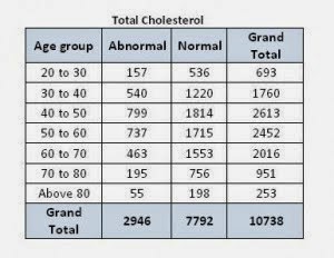
A pan – India survey, conducted by Metropolis Healthcare Ltd ahead of ‘Hypertension Day’ showed a majority of people between 30 to 40 years have alarming LDL cholesterol levels. The survey conducted by Metropolis covered major cities like Delhi, Mumbai, Bangalore and Chennai. Out of the total 10738 lipid profiles done, 25% to 30 % have high Bad Cholesterol (Total Cholesterol, LDL and Triglycerides) and are at a higher risk of acquiring cardiovascular diseases. This could be partly attributed to corporate stress, unhealthy diet and sedentary lifestyle which reflect in the higher LDL cholesterol levels. Also observed was that a fairly substantial number of youngsters examined during the survey have showed the first signs of the onset of a heart ailment — hyper cholesterol and hypertension. 
Individuals have become more aware of the fact that heart disease is not just a disease of the elderly and are now a lot more determined to go a long-way in combating this disease. Calling for regular screening of unhealthy cholesterol and lipid levels, researchers at Metropolis said surveys such as the one conducted would help identify people who are at a greater risk of Hypertension. Preventive measures are required so as to reduce obesity, increasing physical activity, decreasing the salt intake of the population and a concerted effort to promote awareness about hypertension and related risk behaviours. 


The statistics below clearly indicate that young people in the age group 30-40 have high cholesterol, LDL and triglyceride levels and as a result are at a greater risk of hypertension.
by
Dr.Akshaya Srikanth, PharmD., MPH